


Cervical papillomas are one of the manifestations of an infectious disease caused by human papillomavirus. Much depends on the formation of the skin.
Causes of papillomas in the neck
There is one etiological reason why papillomas begin to grow in the neck or anywhere on the human body - infection with human papillomavirus (HPV), a member of the family Papovaviridae. There are more than 100 serotypes of this pathogen, each of which is responsible for the emergence of a different clinical picture of the disease (papilloma, condyloma, warts - these concepts are synonymous, different names are associated with localization features in a particular area).
The main route of infection is the house and genitals in contact (perianal condyloma). The virus can penetrate the skin only in the presence of micro lesions or open wounds, in other cases it can not cross the protective barrier of the skin.
pathogen information
- It is highly prevalent, regardless of gender (more common in women than men), regardless of age or region (according to some reports, 2/3 of the planet is infected with the virus).
- Contains double-stranded, twisted loop DNA that can integrate into the human genome.
- Infection with some strains is associated with a high carcinogenic risk, especially in the case of permanent damage. Cervical papillomas are caused by non-oncogenic strains of the virus.
- The virus goes through two main stages in the division process. In the first stage, it takes an episomal (free) form, and at the same time, the main division of viral particles takes place. This stage is reversible (a long-term remission occurs after treatment). In the second - integrative stage, the virus is implanted in the cell genome (the first step for cell regeneration and the formation of a malignant neoplasm). The first stage is transient and passes relatively quickly, the second explains the presence of latent and carriers.
- The basal layer of the epidermis, where the virus multiplies, is affected. In the remaining layers, the pathogen can persist but cannot divide. As the virus grows in the microbial layer, the normal differentiation of cells in all layers of this region is disrupted, especially at the level of the thorny layer.
- There is a tendency for long-term asymptomatic transport in the body (from a few months to a year). It is rarely possible to determine the moment of a specific infection - this is the reason why treatment is started at the time of intense clinical manifestations, and not at the first vague symptoms.
- Bivalve and quaternary vaccines, which are particularly effective against 16 and 18 most oncogenic strains, are used to prevent infection.
Predisposing factors
- Lack of hygiene. Since the virus can continue its vital activity in the external environment for a long time, it is necessary to carefully follow the rules of personal hygiene when visiting public places (swimming pool, bath, gym).
- Traumatic skin injuries. Micro-cracks or scratches on the skin (for example, caused by rubbing the neck with the collar of a shirt) are enough for the virus to penetrate.
- Immune system disorders. With an immune deficiency of any genesis, favorable conditions for the development of any infection arise. For example, colds and infectious diseases often weaken the immune system and cause papillomas on the skin.
- Do not infect yourself by scratching the skin.
- Systemic lifestyle disorders (stress, lack of physical activity, improper diet). These factors affect the work of all metabolic processes in the body and reduce the barrier function of the skin.
- Environmental factors (hypothermia, excessive ultraviolet exposure) that affect the body's defenses.
External manifestations of the disease
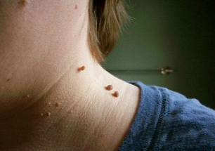
Cervical papillomas look like this in the photo:
- Growth is located at the widest base and extends significantly above the skin surface. Less commonly, the base of the papilloma is represented by a slender foot (in which case the form assumes a hanging position). The second option has a higher risk of injury.
- The boundaries of education are equal and clear.
- The color does not differ from the surrounding skin. In rare cases, it may be fainter or darker than adjacent tissues.
- The surface is often smooth, smooth. Sometimes growths are possible in the upper part of the papilloma, which makes the surface grooved.
- Diameter varies widely - from 1-3 mm to several centimeters (small diameter papillomas are more common).
- Place on any part of the neck (back, front). Sometimes the face gets confused.
As a rule, there are many lesions located along the folds of the skin.
In very rare cases, papillomas in the neck can become malignant, ie degenerate into a skin tumor. This can occur as a result of infection with an oncogenic HPV strain.
The signs that may indicate a malignant transformation are:
- color change and heterogeneity (polymorphism);
- boundary change (blurring, loss of definition);
- view of asymmetry (two equal halves cannot be obtained by drawing a line through the conditional center of the formation);
- intensive growth;
- bleeding or ulceration (a non-specific symptom because it is also typical for simple trauma to the neoplasm);
- itching, burning, peeling;
- dropouts occur (little girl formations around the center).
The appearance of such symptoms does not necessarily mean degeneration of the papilloma, but rather you should learn that we are talking about a common inflammatory mole or skin cancer, consult a doctor and undergo a differential diagnosis.
How to get rid of neck papillomas
Treatment of papillomas in the neck is carried out only in a complex way, simultaneously affecting the pathological focus on the skin and the pathogen itself in the blood.
There are several ways to fight:
Method |
Description |
Medication |
The use of cytostatics, immunomodulators is intended to suppress the replication of the viral agent in the affected area and reduce its concentration in the blood. Some drugs (keratolytics) are applied topically to directly destroy skin growth (causing catarrh and tissue necrosis). |
Physical Methods |
Cryodestruction, laser therapy, electrocoagulation. They aim to get rid of papillomas in both the neck and other parts of the body. These methods allow you to restore the aesthetic appearance of open areas and remove the viral reservoir - the skin neoplasms themselves, but do not completely eliminate the virus from the body. |
Combination therapy |
Combines the two previous options and is therefore the most effective. |
Treatment of papillomas with folk remedies (for example, celandine juice) is ineffective and often dangerous, in any case, one condition is to consult a doctor.
Methods of physical destruction
Formations can be effectively reduced using the following physical methods:
Method |
Description |
Local effects with concentrated acid solutions. |
combination of nitrogen, vinegar, oxalic, lactic acids and copper nitrate trihydrate, etc. A solution of 1. 5% chloropropionate in 50% chloropropionic acid is used, the procedure is performed by a specialist (dermatovenerologist, cosmetologist) in accordance with the rules of surgery. . . . The agent is applied guided with a spatula until the color of the formation becomes clearer (additional application should be stopped immediately as soon as this occurs). For complete cure of papilloma, you need an average of 1-2 treatments. |
Electrocoagulation |
Using a special electric knife, the formation is removed with a dot without affecting the resulting tissues (there is minimal effect on healthy skin cells). The method is most suitable when the formation is a long root and small in size. |
Cryodestruction |
Focus is exposed to liquid nitrogen, which causes ultra-low temperature tissue necrosis. It is good to clear this educational path with a wide base. The duration of nitrogen exposure is selected by a specialist (1-5 minutes). After moxibustion, a burn develops that improves within an average of 10 days. |
Laser removal |
The most modern and sensitive approach to eliminate growths in prominent places such as the neck. Has the most positive feedback. In continuous mode, they move in focus with the help of a light guide for 5 seconds to 3 minutes. The healing time is very short compared to other methods (5-7 days). The technique is associated with minimal trauma to surrounding tissues due to high accuracy of movement. |
Classical surgery (excision with a scalpel) |
Very rarely, it is used only with large lesions or suspected malignancy. The reason is that the lesions are often large, scattered around the neck and too small for excision, in addition, after surgical excision, they may leave scars that create a cosmetic defect. |